Home > Products > Tumor Cell Lines > Human Tumor Cell Lines >

MDA-MB-453

Product Name

MDA-MB-453

Price Get Quote
Product Code

MDA-MB 453; MDA MB 453; MDA-MB453; MDAMB453; MDA-453; MDA453; MD Anderson-Metastatic Breast-453

Cat.No

ABC-TC0656

Species

Human

Size/Quantity

1 vial

Biosafety Level

1

Shipping Info

Dry Ice

Storage

Liquid Nitrogen

Description

MDA-MB-453 cells were derived in 1976 from a female patient with metastatic breast cancer. These cells have a deletion mutation in the p53 gene, resulting in the absence of the p53 protein. They exhibit aneuploidy, with abnormal numbers of chromosomes, including the absence or under-representation of certain normal chromosomes. Marker chromosomes, with structural abnormalities, are also present. Over time, the cell population has transitioned from near-diploid to tetraploid. MDA-MB-453 cells overexpress fibroblast growth factor (FGF) receptors. Overall, these cells represent a breast cancer cell line with distinct genetic, chromosomal, and receptor expression characteristics.

Why choose MDA-MB-453 from AcceGen?

Our MDA-MB-453 cell product is sterile, being negative for bacterial, fungal, mycoplasma, and human pathogen contamination. It ensures high quality, viability, and adheres to stringent production standards. The cells exhibit a healthy morphology, and their identity is confirmed using Short Tandem Repeat (STR) analysis.

Source Organ

Breast

Recommended Medium And Supplement
Citation Guide

When you publish your research, please cite our product as “AcceGen Biotech Cat.# XXX-0000”. In return, we’ll give you a $100 coupon. Simply click here and submit your paper’s PubMed ID (PMID).

Application

FOR RESEARCH USE ONLY

MDA-MB-453 cells have diverse applications in breast cancer research. They are used to examine the anti-proliferative effects of drugs and uncover the molecular mechanisms behind therapeutic efficacy. Additionally, these cells aid in studying tumorigenesis, endocrine resistance signaling, and identifying potential markers for breast cancer. By assessing drug responses, investigating tumor pathways, and analyzing gene and protein profiles, MDA-MB-453 cells contribute to understanding breast cancer biology and advancing targeted therapies. Their versatility makes them valuable tools in research aiming to improve treatment outcomes for breast cancer patients.

Growth Conditions

37 ℃

Cell Type

Epithelial

Growth Mode

Adherent

Product Type

Human Breast Cancer Cell Lines

Product Image

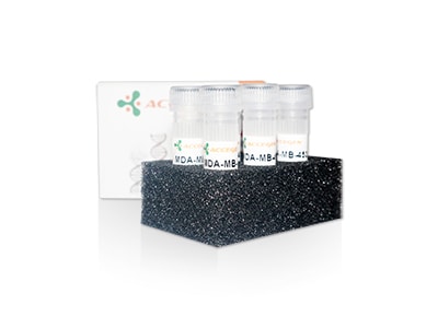
mda-mb-453 cell line

AcceGen product mda-mb-453 cell line

Frequently Asked Questions

  • What are MDA-MB-453 cells, and why should I use them in my research?

    MDA-MB-453 cells are a human breast cancer cell line that originates from a pleural effusion of a patient with metastatic breast cancer. These cells are often used in cancer research.

  • What specific growth factors or supplements can enhance the activation of MDA-MB-453 cells?

    Enhancing the activation and growth of MDA-MB-453 cells can be achieved by using specific growth factors and supplements in the culture medium. Here are some key factors and supplements that can be used:

    1. Growth Factors
    Epidermal Growth Factor (EGF): EGF is known to stimulate cell growth and proliferation. Adding EGF to the culture medium can enhance the growth of MDA-MB-453 cells. Typical concentration: 10-20 ng/mL.

    Insulin-like Growth Factor 1 (IGF-1): IGF-1 plays a significant role in cell growth and survival. It can be used to promote the proliferation of breast cancer cells, including MDA-MB-453. Typical concentration: 10-50 ng/mL.

    Heregulin (HRG): Heregulin is a ligand for the HER3 receptor, which can activate HER2 and HER3 signaling pathways. It can be used to enhance growth and signaling in HER2-positive cells. Typical concentration: 10-50 ng/mL.

    2. Hormones
    Androgens (e.g., Dihydrotestosterone, DHT): Since MDA-MB-453 cells express the androgen receptor (AR), adding androgens like dihydrotestosterone (DHT) can activate AR signaling and influence cell growth. Typical concentration: 1-10 nM.

    3. Supplements
    Insulin: Insulin is often added to cell culture media to promote growth and enhance glucose uptake. Typical concentration: 10 µg/mL.

    Hydrocortisone: Hydrocortisone can be used to enhance the growth of certain breast cancer cell lines, including those expressing steroid hormone receptors. Typical concentration: 1 µg/mL.

    Fetal Bovine Serum (FBS): FBS is a common supplement in cell culture media that provides essential growth factors, hormones, and nutrients.Typical concentration: 10-20%.

  • How do I passage MDA-MB-453 cells?

    Procedure

    Preparation: Warm the RPMI-1640 medium, PBS, and trypsin-EDTA solution to 37°C in a water bath. Ensure all necessary equipment and reagents are sterile and ready for use.

    Observe Cells: Place the flask containing MDA-MB-453 cells under an inverted microscope.
    Check for cell confluence (typically, cells should be 70-80% confluent before passaging).

    Remove Old Medium: In a biosafety cabinet, aspirate the old medium from the flask carefully without disturbing the cell monolayer.

    Rinse Cells: Add 5-10 mL of PBS to the flask to rinse off any residual medium and serum that may inhibit trypsin activity. Gently rock the flask to ensure the cells are thoroughly rinsed, then aspirate the PBS.

    Add Trypsin-EDTA: Add enough trypsin-EDTA solution to cover the cell monolayer (typically 1-2 mL for a T-75 flask). Gently tilt the flask to ensure the entire surface is covered with trypsin-EDTA.

    Incubate: Place the flask in the incubator for 3-5 minutes, checking the cells under the microscope periodically. The cells should round up and detach from the surface.

    Neutralize Trypsin: Once the cells are detached, add 5-10mL of complete RPMI-1640 medium (with FBS) to the flask to neutralize the trypsin.

    Collect Cells: Gently pipette the cell suspension up and down to ensure all cells are detached and in suspension. Transfer the cell suspension to a 15mL conical tube.

    Centrifuge: Centrifuge the cell suspension at 200×g for 5 minutes to pellet the cells.

    Resuspend Cells: Carefully aspirate the supernatant without disturbing the cell pellet. Resuspend the cells in an appropriate volume of fresh complete RPMI-1640 medium.

    Count Cells (Optional): If needed, take a small aliquot of the cell suspension to count cells using a hemocytometer or an automated cell counter.

    Seed New Flasks: Seed the cells into new culture flasks or plates at the desired density. For example, if splitting at a 1:5 ratio, add 1 part of the cell suspension to 4 parts of fresh medium in a new flask.

    Incubate: Place the newly seeded flasks or plates back into the incubator set at 37°C with 5% CO2.

    Monitoring: Observe the cells under the microscope after 24 hours to ensure they have adhered and are spreading out. Change the medium every 2-3 days or as needed.

  • ONLINE INQUIRY
  • PRODUCT REVIEWS
We know how valuable your research is to you, but are you wondering what you can expect to pay for quick accurate results every time?

Fill out a request in the form below and we’ll get back to you within 24 hours with a quote.
Privacy Policy: AcceGen will never sell, rent, or share your personal information with any third parties without your express permission.
Reviews of MDA-MB-453
AcceGen is always trying to do right by our customers and working hard to build a higher quality product.

Your email address will not be published.

AcceGen Scroll Top Button