1
Discover top-quality products tailored for scientific and medical research. Request a personalized quote today
to enhance your projects.
Product Code | MHSteC |
Species | Mouse |
Cat.No | ABC-TC3927 |
Quality Control | All cells test negative for mycoplasma, bacteria, yeast, and fungi. |
Product Category | Primary Cells |
Size/Quantity | 1 vial |
Cell Type | Stellate Cell |
Shipping Info | Dry Ice |
Growth Conditions | 37 ℃, 5% CO2 |
Source Organ | Liver |
Disease | Normal |
Biosafety Level | 1 |
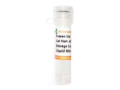
Storage | Liquid Nitrogen |
Product Type | Mouse Primary Cells |
Mouse Hepatic Stellate Cells (MHSCs) are primary mesenchymal cells isolated from mouse liver tissue. Primary MHSCs demonstrate limited proliferative capacity and cryopreserved immediately after isolation. These cells reside in the perisinusoidal space of Disse and account for 5–8% of hepatic cells. These cells derived from bone marrow precursors and store up to 80% of the total body vitamin A. Upon liver injury or stress, MHSCs transition into activated myofibroblasts, characterized by increased proliferation and extracellular matrix (ECM) secretion, driving fibrotic responses in conditions like non-alcoholic steatohepatitis and toxin-induced cirrhosis. Primary MHSCs retain physiological vitamin A storage and ECM remodeling functions, making them ideal for modeling early fibrotic pathways.
When you publish your research, please cite our product as “AcceGen Biotech Cat.# XXX-0000”. In return, we’ll give you a $100 coupon. Simply click here and submit your paper’s PubMed ID (PMID).
Mouse Hepatic Stellate Cells offer a robust model system for delving into various facets of liver metabolism and pathophysiology. Their application spans diverse cell-based assays, including toxicity assessment, drug screening, and metabolic studies. They emerge as a crucial resource in the study of liver fibrosis, offering insights that could drive innovative therapeutic strategies for hepatic fibrosis treatment, ultimately aiming to enhance patient outcomes and mitigate the impact of chronic liver injury.
These cells become activated during liver injury, contributing to fibrosis and scar tissue formation.
In some cases, with appropriate signals, they can return to a less active, quiescent state, though this is limited.
They interact closely with hepatocytes and immune cells, influencing inflammation and tissue remodeling.
Key pathways include TGF-β, PDGF, and Hedgehog signaling, which drive the activation and proliferation of these cells.