- In-Stock Tumor Cell Lines
- Human Orbital Fibroblasts
- Human Microglia
- Human Pulmonary Alveolar Epithelial Cells
- Human Colonic Fibroblasts
- Human Type II Alveolar Epithelial Cells
- Human Valvular Interstitial Cells
- Human Thyroid Epithelial Cells
- C57BL/6 Mouse Dermal Fibroblasts
- Human Alveolar Macrophages
- Human Dermal Fibroblasts, Adult
- Human Lung Fibroblasts, Adult
- Human Retinal Muller Cells
- Human Articular Chondrocytes
- Human Retinal Pigment Epithelial Cells
- Human Pancreatic Islets of Langerhans Cells
- Human Kidney Podocyte Cells
- Human Renal Proximal Tubule Cells
Function of Luciferase Reporter Cell Lines
Luciferase reporter cell lines utilize luciferase expression as a bioluminescent marker to monitor specific biological processes. When a luciferase-specific substrate is introduced, the enzyme catalyzes a light-producing reaction, enabling researchers to visualize and quantitatively measure gene expression, signaling pathway activation, or protein-protein interactions. This system provides a high signal-to-noise ratio, which is particularly advantageous in drug screening and mechanistic studies of disease.
In these engineered cell lines, the luciferase gene is placed under the control of a defined promoter, response element, or fused to a gene of interest. Activation of the regulatory element by transcription factors or signaling molecules leads to transcription of luciferase mRNA and subsequent translation into the functional enzyme. The luciferase enzyme then catalyzes its substrate, releasing detectable photons. The emitted bioluminescence, measured by instruments such as luminometers, correlates directly with the activity of the pathway under investigation.
Owing to their high sensitivity and dynamic range, luciferase reporter systems are widely employed in gene expression analysis, signal transduction research, and drug discovery. They permit detection of subtle transcriptional changes and yield reproducible data suitable for downstream validation. Furthermore, these reporter systems are adaptable to various experimental formats, including cell-based assays, three-dimensional culture models, and in vivo imaging. Essentially, luciferase serves as a reporter gene that converts a molecular event into a quantifiable optical signal.
Flow Cytometry Detection of Immunostained Luciferase Reporter Cells
We have developed a flow cytometry-based method to detect luciferase reporter expression at the single-cell level. This approach utilizes immunostaining with an antibody specific for the Luc1 variant of luciferase in the C4-2B Luc1 stable cell line. The assay was optimized and validated to ensure robust detection of luciferase protein. By calculating the fold-change in fluorescence intensity relative to control cells, this method achieves high precision and reproducibility. Consequently, it allows accurate, high-throughput quantification of reporter expression across individual cells within a population.
Experimental Section
Aim
To detect the expressed positive cell population of Luciferase in C4-2B Luc1 cells by flow cytometry.
Materials and Reagents
| Name | Manufacture | Cat# | Lot# |
|---|---|---|---|
| dPBS | Gibco | 10010-023 500ML | 3013794 |
| HI FBS Heat Inactivated Fetal Bovine Serum | Gibco | 10438-026 | 2535232RP |
| Human TruStain FcX™ (Fc Receptor Blocking Solution) 200X | Biolegend | 422302 | B443035 |
| Aqua fluorescent reactive dye | Invetrogen | L34966A | 2983213 |
| FOXP3 Fixation/Permeabilization Buffer(100-test) | Biolegend | 421403 | B435799 |
| Firefly luciferase Recombinant Rabbit Monoclonal Antibody (HL2164) | ThermoFisher | MA550410 | 79704002 |
| Goat anti-Rabbit IgG (Heavy chain) Superclonal™ Secondary Antibody, Alexa Fluor™ Plus 647 | Scientific | A5505550UL | 3318841 |
| 96-well u-bottom plate | LabForce | 210AAM447 | 022824A001 |
| Cell info | Testing for |
|---|---|
| C4-2B Luc1 | Testing cell line for Luc1 expression |
Instrument
Invitrogen™, Attune™ NxT Flow Cytometer
Procedure
1. Collect all cells into a 15 mL centrifuge.
2. Spin cells at 300g for 5 min.
3. Resuspend cells with dPBS.
4. Count cell density.
5. Load 1.0E+06 cells/well of 96-well plate per each cell line detailed in the plate map shown below.
| Cell | Staining info |
|---|---|
| C4-2B Luc1 | Live/Dead staining only |
| C4-2B Luc1 | AF-647-2nd antibody only |
| C4-2B Luc1 | Stain for Luc1 + AF-647-2nd antibody |
6. Spin cells at 300g for 5 min.
7. Wash cells once with cold-dPBS.
8. Spin cells at 300g for 5 min.
9. Resuspend cells in Live/Dead solution except the cells for No staining detailed below.
| Reagent | Dilution | Volume (μL)/Each |
|---|---|---|
| Aqua fluorescent reactive dye | 1000X | 0.1 |
| dPBS | 99.9 |
10. Incubate at 4°C for 15 min.
11. Add 150 μL ddPBS/well.
12. Spin cells at 300g for 5 min.
13. Remove the supernatant.
14. Add 1X working concentration of Fix/Perm 200 μL/well.
15. Incubate at room temperature for 20 min in dark.
16. Spin cells at 300g for 5 min.
17. Wash cells with dPBS at 200 μL/well.
18. Spin cells at 300g for 5 min.
19. Wash cells once with 1X Perm buffer at 200 μL/well.
20. Spin cells at 300g for 5 min.
21. Add diluted Human TruStain FcX™ (Fc Receptor Blocking Solution) 100 μL/well.
22. Incubate at 4°C for 15 min.
23. Spin cells at 300g for 5 min.
24. Resuspend cells in 100 μL of diluted either anti-Firefly Luciferase Antibody solution or dPBS (for IgG control antibody only) per well (dilution factor shown below).
| Manufacture | Cat# | Dilution | |
|---|---|---|---|
| Firefly luciferase Recombinant Rabbit Monoclonal Antibody | ThermoFisher Scientific | MA550410 | 1:500 |
| Goat anti-Rabbit IgG (Heavy chain) Superclonal™ Secondary Antibody, Alexa Fluor™ Plus 647 | ThermoFisher Scientific | A5505550UL | 1:1000 |
25. Incubate at 4°C for 30 min in dark.
26. Add 100 μL cold dPBS/well.
27 . Spin cells at 300g for 5 min.
28. Resuspend cells in diluted Goat anti-Rabbit IgG (Heavy chain) Superclonal™ Secondary Antibody solution at 100 μL/well.
30. Incubate at 4°C for 20-30 min in dark.
31. Add 100 μL cold dPBS/well.
32. Spin cells at 300g for 5 min.
33. Wash cells with dPBS once with keeping in dark.
34. Spin cells at 300g for 5 min.
35. Resuspend cells in dPBS with keeping in dark, following running flow on flow cytometry, otherwise resuspend cells in 1X flow buffer with keeping in dark. Keep the cells at 4°C for flow.
Flow Data Report
Gating info
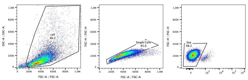
Flow Data

Result
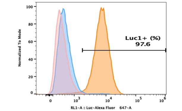
1. The anti-Firefly Luciferase Antibody exhibits specific binding to luciferase expression in C4-2B Luc1 cells.
2. 97.6% of the C4-2B Luc1 cell population shows positive Luc1 expression.
Conclusion
Luciferase reporters remain a widely utilized tool in biological research due to their convenience, sensitivity, and suitability for high-throughput applications. A broad selection of luciferase-expressing reporter cell lines, including the C4-2B Luc1 line described here, is available through AcceGen. The flow cytometric assay presented is not only applicable to Luc1-based cell lines but can also be extended to other luciferase reporter systems. Although the current antibody does not recognize the Luc2 variant, future incorporation of antibodies targeting additional luciferase isoforms, such as Luc2, would further broaden the utility of this approach. Such development would enhance the applicability of flow cytometry for quality control and functional validation of diverse luciferase reporter cell lines.
Copyright - Unless otherwise stated all contents of this website are AcceGen™ All Rights Reserved – Full details of the use of materials on this site please refer to AcceGen Editorial Policy – Guest Posts are welcome, by submitting a guest post to AcceGen you are agree to the AcceGen Guest Post Agreement – Any concerns please contact marketing@accegen.com