1
Discover top-quality products tailored for scientific and medical research. Request a personalized quote today
to enhance your projects.
Product Code | HCEpiC |
Species | Human |
Cat.No | ABC-TC3577 |
Quality Control | All cells test negative for mycoplasma, bacteria, yeast, and fungi. |
Product Category | Primary Cells |
Size/Quantity | 1 vial |
Cell Type | Epithelial |
Shipping Info | Dry Ice |
Growth Conditions | 37 ℃, 5% CO2 |
Source Organ | Corneal |
Disease | Normal |
Biosafety Level | 1 |
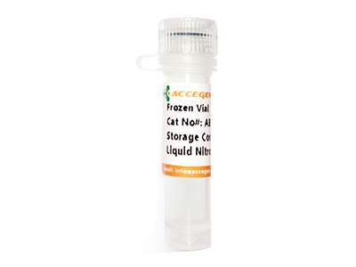
Storage | Liquid Nitrogen |
Product Type | Ocular Cells |
Key Features | -Backed by AcceGen advanced technology |
Human Corneal Epithelial Cells originate from the outermost layer of the cornea, the stratified squamous epithelium, which can be divided into three layers: basal cells, wing cells, and surface squamous cells . They express cytokeratin 3 and cytokeratin 12, markers specific to differentiated corneal epithelial cells, detectable by immunocytochemistry.Their morphology shows typical polygonal squamous epithelial-like features. These cells can form a mechanical barrier to resist the invasion of pathogens, secrete antimicrobial peptides (such as defensins) and cytokines to participate in innate immunity.Dysfunction of these cells is associated with diseases such as dry eye syndrome, infectious keratitis, and corneal epithelial defects. In addition, they can maintain corneal transparency through tight junctions.
When you publish your research, please cite our product as “AcceGen Biotech Cat.# XXX-0000”. In return, we’ll give you a $100 coupon. Simply click here and submit your paper’s PubMed ID (PMID).
Human Corneal Epithelial Cells can be used to study corneal epithelial barrier function, wound healing mechanisms, and innate immune responses, such as studying the interaction between pathogens and the cornea through in vitro models. These cells can also be used to evaluate the permeability, toxicity, and repair effects of ocular drugs (such as antibiotics and anti-inflammatory drugs).