1
Discover top-quality products tailored for scientific and medical research. Request a personalized quote today
to enhance your projects.
Product Code | T3M4; Panc89; Panc-89; PANC-89; Panc 89 |
Species | Human |
Cat.No | ABC-TC1324 |
Product Category | Tumor Cell Lines |
Size/Quantity | 1 vial |
Shipping Info | Dry Ice |
Growth Conditions | 37 ℃, 5% CO2 |
Biosafety Level | 1 |
Storage | Liquid Nitrogen |
Product Type | Human Pancreas Cancer Cell Lines |
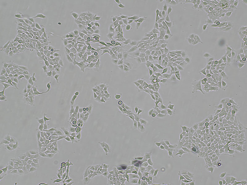
T3M-4 is a human pancreatic adenocarcinoma cell line originally derived from a primary pancreatic exocrine tumor and established through xenografting into athymic nude mice. The cells exhibit epithelial morphology and typically grow as an adherent monolayer. Morphologically, T3M-4 cells show epithelial-like features under standard conditions, while co-culture with activated CD4+ T cells induces a more spindle-shaped phenotype. Chromosomal analysis confirms a human hyperdiploid karyotype. Genetically, T3M-4 cells harbor mutations in KRAS (Q61H) and TP53, both commonly implicated in pancreatic cancer. They are known to secrete carcinoembryonic antigen (CEA) both in vitro and in vivo. T3M-4 cells are tumorigenic when transplanted into nude mice and retain their histological characteristics upon serial passage. The line originates from a lymph node metastasis of pancreatic cancer, indicating metastatic potential.
When you publish your research, please cite our product as “AcceGen Biotech Cat.# XXX-0000”. In return, we’ll give you a $100 coupon. Simply click here and submit your paper’s PubMed ID (PMID).
The T3M-4 cell line offers multiple applications in the field of human pancreatic cancer research. Firstly, it can be used to identify the gene profile of human pancreas carcinoma, supporting analysis of oncogene activation as well as aberrant expression and epigenetic changes of genes specific to pancreatic cancer. This aids in understanding the molecular mechanisms underlying the disease. Additionally, T3M-4 cells serve as a useful tool for evaluating the anti-tumor potential of antibodies or drugs, aiding the development of new therapeutic strategies for treating human pancreatic cancer. By assessing the efficacy of different treatments, researchers can advance the search for effective therapies targeting this challenging malignancy.
Approximately 1-2 ×10^4 cells/cm².
Insufficient Trypsinization: The cells may not have been trypsinized long enough, leading to incomplete detachment and clumping.
Excessive Trypsinization: Prolonged exposure to trypsin can damage cell surface proteins, affecting their ability to reattach.
Incorrect Trypsin Concentration: Using a trypsin solution that is too concentrated can harm the cells, while too dilute may not be effective.
Incomplete Neutralization of Trypsin: If trypsin is not completely neutralized with serum-containing medium after detachment, residual trypsin can continue to damage cells.
Cell Density: Plating cells at too high or too low a density can affect their ability to attach and spread out.
Suboptimal Culture Conditions: Temperature, pH, or medium composition might not be optimal for cell attachment.
Optimize Trypsinization Time: Ensure cells are exposed to trypsin-EDTA for an appropriate duration (usually 2-5 minutes at 37°C) to detach without excessive damage. Monitor under a microscope.
Neutralize Trypsin Effectively: Immediately add an appropriate amount of medium containing serum to neutralize the trypsin after detachment.
Use Gentle Pipetting: Gently pipette the cells up and down to break up clumps without causing excessive mechanical damage.
Optimize Seeding Density: Seed cells at an appropriate density (e.g., 1-2 x 10^4 cells/cm²) to allow for optimal attachment and growth.
Check Culture Conditions: Ensure the culture medium is pre-warmed to 37°C and that the CO₂ levels and pH are appropriate.
Coating Culture Vessels: If cells still do not attach well, consider coating the culture vessels with extracellular matrix proteins (e.g., collagen, fibronectin) to enhance cell adhesion.