- In-Stock Tumor Cell Lines
- Human Orbital Fibroblasts
- Human Microglia
- Human Pulmonary Alveolar Epithelial Cells
- Human Colonic Fibroblasts
- Human Type II Alveolar Epithelial Cells
- Human Valvular Interstitial Cells
- Human Thyroid Epithelial Cells
- C57BL/6 Mouse Dermal Fibroblasts
- Human Alveolar Macrophages
- Human Dermal Fibroblasts, Adult
- Human Lung Fibroblasts, Adult
- Human Retinal Muller Cells
- Human Articular Chondrocytes
- Human Retinal Pigment Epithelial Cells
- Human Pancreatic Islets of Langerhans Cells
- Human Kidney Podocyte Cells
- Human Renal Proximal Tubule Cells
Reporter cell lines are common stable cell lines that have been labeled with a reporter gene, such as luciferase, Green fluorescent protein (GFP), Red fluorescent protein (RFP), and so on. Researchers use reporter gene assays to study signaling pathways, gene regulation, and the structure of regulatory elements. Luciferase reporter cell lines provide a highly sensitive and simple way for researchers.
How to use luciferase?
As a reporter gene, the luciferase gene is inserted into a lentiviral vector and then integrated into the chromosome of the cell genome. When the cells proliferate, the progeny cells will also express the luciferase gene. The luciferase gene can be expressed stably and continuously in the cells[1, 2].
Luciferin will be catalyzed into oxyluciferin by the luciferase when oxygen and Adenosine triphosphate (ATP) are present. And fluorescence occurs during this reaction.【Figure 1】
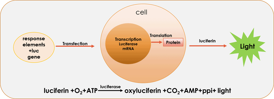
Figure 1. The catalytic reaction of luciferase
Dual-Luciferase reporter system
Dual-Luciferase reporter gene assay system is commonly used in scientific research, which can express both Firefly luciferase (FLuc) and Renilla luciferase (RLuc) in cells. FLuc is used to report the expression of the gene of interest, and RLuc is used to report the internal reference gene[3]. Both of them can catalyze their own substrates to emit different fluorescence and do not interfere with each other during fluorescence detection. Then the fluorescence intensity is detected by the instrument to obtain the expression level of the gene [5][Figure 2]. In this way, the influence of factors such as cell viability and transfection efficiency on the experiment can be minimized, and the reliability of the experiment can be increased[4].
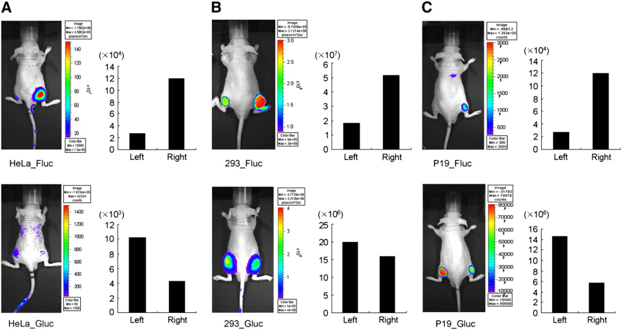
Figure 2. In vivo visualization of expression of dual-luciferase in mice [5]
Luciferase reporter cell lines has a wide range of applications [6]. For example, the growth of tumor cells, trafficking of immune cells, migration of transplanted cells, and so on. In addition, it will be quick and accurate to research the disease process and develop new cell therapies by establishing animal models [7-10].
Tumor cell detecting
A lot of studies have employed Luciferase as a reporter gene in real-time in vivo imaging. Researchers can directly and quickly detect tumor growth and migration by injecting the luciferase reporter cell line into experimental animals. This tumor animal model provides a powerful tool for the research of tumorigenesis mechanisms[11-13].
Immune cell tracing
Researchers can observe the identification and killing of pathogens and tumor cells by luciferase reporter immune cells. And they can track the proliferation and migration of immune cells.
Stem cell tracing
Researchers can track the proliferation, differentiation and migration of stem cells in living animals by transplanting luciferase reported stem cells into animals[14-16].
Research for drug development
Scientists use luciferase cell lines in the development and research of anti-tumor drugs. They transplanted the luciferase tumor cell line into experimental animals, which can provide useful information for formulating the administration time, dosage and route of administration of antitumor drugs[12, 17]. In pharmacology, researchers mark genes of interest with luciferase genes to create the transgenic mice for observing drug effects, which has been widely used in tumor research[18-21].
Research on gene expression and promoter
Researchers insert the luciferase gene into the downstream of the promoter of the gene of interest to realize the co-expression of luciferase and the target gene, so that the expression of the target gene can be directly observed. Dual-luciferase assays can detect promoter activity rapidly and accurately and facilitate the analysis of promoter structure. Luciferase has been widely used in cell lines now[18, 22].
A Great Tool For Researchers
Luciferase has shown great potential in cell tracing, drug development and gene therapy. With the advantages of high sensitivity and accurate positioning, luciferase reporter cell line has become a powerful tool for researchers to study in vitro luminescence assay and in vivo bioluminescence imaging. Now it has been widely used in laboratory and clinical research. We look forward to more diversified applications of luciferase in the future.
Where to Get Luciferase Reporter Cell Line for Your Research?
AcceGen offers reporter stable cell lines for almost any application regardless of cell type with lentiviral-based transduction. Responsive reporter cells form the core of cell-based assays for screening and identifying drugs, antibodies, cell factors, and other effectors. These cell lines provide you with a convenient means to research. To get more information, please refer to: Reporter Stable Cell Lines.
It is our pleasure to help relative researches to move forward. All the products of AcceGen strictly comply with international standards. For more detailed information, please visit our product portfolio or contact inquiry@accegen.com.
References
1. Li, S., et al., Recent achievements of bioluminescence imaging based on firefly luciferin-luciferase system. Eur J Med Chem, 2021. 211: p. 113111.
2. Smale, S.T., Luciferase assay. Cold Spring Harb Protoc, 2010. 2010(5): p. pdb prot5421.
3. Xu, Y.Z., et al., Promoter deletion analysis using a dual-luciferase reporter system. Methods Mol Biol, 2013. 977: p. 79-93.
4. Kenda, M., et al., Evaluation of Firefly and Renilla Luciferase Inhibition in Reporter-Gene Assays: A Case of Isoflavonoids. Int J Mol Sci, 2021. 22(13).
5. Lee, J.Y., et al., Development of a dual-luciferase reporter system for in vivo visualization of MicroRNA biogenesis and posttranscriptional regulation. J Nucl Med, 2008. 49(2): p. 285-94.
6. Kim, S.B. and R. Paulmurugan, Bioluminescent Imaging Systems for Assay Developments. Anal Sci, 2021. 37(2): p. 233-247.
7. Liu, S., et al., Brightening up Biology: Advances in Luciferase Systems for in Vivo Imaging. ACS Chem Biol, 2021. 16(12): p. 2707-2718.
8. Viviani, V.R., G.F. Pelentir, and V.R. Bevilaqua, Bioluminescence Color-Tuning Firefly Luciferases: Engineering and Prospects for Real-Time Intracellular pH Imaging and Heavy Metal Biosensing. Biosensors (Basel), 2022. 12(6).
9. Syed, A.J. and J.C. Anderson, Applications of bioluminescence in biotechnology and beyond. Chem Soc Rev, 2021. 50(9): p. 5668-5705.
10. Foucault, M.L., et al., In vivo bioluminescence imaging for the study of intestinal colonization by Escherichia coli in mice. Appl Environ Microbiol, 2010. 76(1): p. 264-74.
11. Zhu, L., et al., Targeting and Therapy of Glioblastoma in a Mouse Model Using Exosomes Derived From Natural Killer Cells. Front Immunol, 2018. 9: p. 824.
12. Iwano, S., et al., Single-cell bioluminescence imaging of deep tissue in freely moving animals. Science, 2018. 359(6378): p. 935-939.
13. Trinh, T.L., et al., Immune evasion by TGFbeta-induced miR-183 repression of MICA/B expression in human lung tumor cells. Oncoimmunology, 2019. 8(4): p. e1557372.
14. Peng, Y., et al., Generation of a luciferase-expressing human embryonic stem cell line: NERCe002-A-2. Stem Cell Res, 2018. 28: p. 172-176.
15. Han, D. and J.C. Wu, Using Bioengineered Bioluminescence to Track Stem Cell Transplantation In Vivo. Methods Mol Biol, 2020. 2126: p. 1-11.
16. Conway, M., et al., Real-time tracking of stem cell viability, proliferation, and differentiation with autonomous bioluminescence imaging. BMC Biol, 2020. 18(1): p. 79.
17. Roberts, R.S., et al., Renilla luciferase as a reporter to assess SARS-CoV mRNA transcription regulation and efficacy of anti-SARS-CoV agents. Adv Exp Med Biol, 2006. 581: p. 597-600.
18. Li, Q., H. Yoshimura, and T. Ozawa, A Split-Luciferase-Based Cell Fusion Assay for Evaluating the Myogenesis-Promoting Effects of Bioactive Molecules. Methods Mol Biol, 2021. 2274: p. 79-87.
19. Roda, A., et al., A new gastric-emptying mouse model based on in vivo non-invasive bioluminescence imaging. Neurogastroenterol Motil, 2010. 22(10): p. 1117-e288.
20. Kim, S.J., et al., Homogeneous Immunoassay Using a Tri-Part Split-Luciferase for Rapid Quantification of Anti-TNF Therapeutic Antibodies. ACS Sens, 2021. 6(5): p. 1807-1814.
21. Hoenen, T., Luciferase-Expressing Ebolaviruses as Tools for Screening of Antivirals. Methods Mol Biol, 2017. 1628: p. 189-194.
22. Alcaraz-Perez, F., V. Mulero, and M.L. Cayuela, Application of the dual-luciferase reporter assay to the analysis of promoter activity in Zebrafish embryos. BMC Biotechnol, 2008. 8: p. 81.
Copyright - Unless otherwise stated all contents of this website are AcceGen™ All Rights Reserved – Full details of the use of materials on this site please refer to AcceGen Editorial Policy – Guest Posts are welcome, by submitting a guest post to AcceGen you are agree to the AcceGen Guest Post Agreement – Any concerns please contact marketing@accegen.com