- In-Stock Tumor Cell Lines
- Human Orbital Fibroblasts
- Human Microglia
- Human Pulmonary Alveolar Epithelial Cells
- Human Colonic Fibroblasts
- Human Type II Alveolar Epithelial Cells
- Human Valvular Interstitial Cells
- Human Thyroid Epithelial Cells
- C57BL/6 Mouse Dermal Fibroblasts
- Human Alveolar Macrophages
- Human Dermal Fibroblasts, Adult
- Human Lung Fibroblasts, Adult
- Human Retinal Muller Cells
- Human Articular Chondrocytes
- Human Retinal Pigment Epithelial Cells
- Human Pancreatic Islets of Langerhans Cells
- Human Kidney Podocyte Cells
- Human Renal Proximal Tubule Cells
The Endocrine System
The endocrine system constitutes a complex, highly integrated network of glands and organs responsible for the synthesis and secretion of signaling biomolecules known as hormones. These hormones are released into the bloodstream or interstitial fluid to regulate physiological processes throughout the body, functioning as one of the two primary signaling networks alongside the nervous system. Hormones serve as the principal messengers of this system, traveling via the circulatory system to engage specific target cells and organs, thereby modulating a diverse array of functions including metabolism, growth and development, homeostasis, sexual function, reproduction, and circadian rhythms [1]. Despite their minute concentrations, hormones are capable of eliciting profound physiological responses and are indispensable for life and health.
Glands and Organs of the Endocrine System
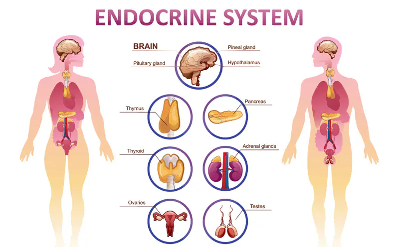
The endocrine system is primarily composed of specialized glands and organs including the hypothalamus, pituitary gland, pineal gland, thyroid and parathyroid glands, adrenal glands, thymus, pancreas, and the gonads (ovaries and testes), as illustrated in Figure 1 [1]. In addition, adipose tissue is now recognized as a vital, active endocrine organ rather than merely a passive energy storage site. Each of these tissues synthesizes distinct hormones with specific physiological effects.
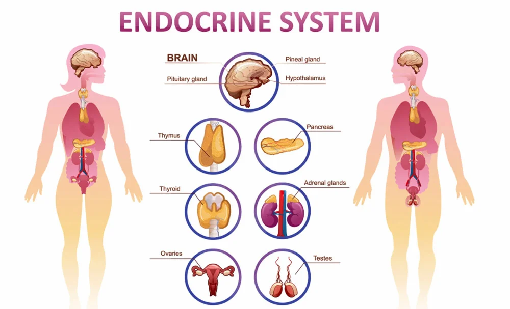
Figure 1. The Endocrine System (Cited from The Endocrine Society).
The hypothalamus serves as the primary coordinating center, producing hormones such as anti-diuretic hormone (ADH), corticotropin-releasing hormone (CRH), gonadotropin-releasing hormone (GnRH), growth hormone-releasing hormone (GHRH), growth hormone-inhibiting hormone (GHIH), prolactin-releasing hormone (PRH), prolactin-inhibiting hormone, (PIH), and thyrotropin-releasing hormone (TRH).
The pituitary gland, a pea-sized structure located at the base of the brain inferior to the hypothalamus, secretes hormones via its anterior and posterior lobes that influence growth, metabolism, reproduction, and stress response [2]. There are eight hormones released by the pituitary gland: gonadotropins (LH and FSH), growth hormone (GH), thyroid stimulating-hormone (TSH), adrenocorticotropic hormone (ACTH), prolactin, ADH and oxytocin.
The pineal gland is a small and pinecone-shaped organ located at the posterior of the diencephalon region in the brain. It secretes melatonin in response to the light impulse, thereby regulating the sleep-wake cycles and other circadian rhythms.
The thyroid is also called the thyroid gland. It sits in the anterior neck just inferior to the larynx, served by large arteries with many branches and a dense network of capillaries. The parathyroid glands are four tiny circular glandular structures embedded in the posterior surface of the thyroid gland. The thyroid and parathyroid glands regulate many hormonal systems. They produce thyroid hormones thyroxine (T4), triiodothyronine (T3), calcitonin and parathyroid hormone (PTH), which are critical to the healthy development and maturation of vertebrates and metabolism regulation.
The adrenal glands are located superior to each kidney, and function synergistically with the hypothalamus and pituitary gland. They make and release corticosteroid hormones and epinephrine which are vital for stress response, blood pressure, and metabolism.
The thymus is located in the upper thoracic region. It functions as an endocrine gland by secreting hormones such as thymopoietin, thymosin, thymulin, and thymic humoral factor, which are crucial for T-cell maturation and immune system development.
The pancreas is primarily a digestive organ. It excretes pancreatic juice into the small intestine via the pancreatic duct. The pancreas contains individual islets of Langerhans which make up less than 2% of pancreatic tissue, and are responsible for producing glucagon and insulin, both of which help regulate the glucose concentration in the blood.
Furthermore, adipose tissue is now recognized as a metabolically active endocrine organ. It is a connective tissue that distributes throughout the body (subcutaneous, visceral, and bone marrow). Adipose tissue releases many different hormones, including leptin, angiotensin, adiponectin, and oestrogen.
Endocrine System Disorders
The precise functioning of the endocrine system is fundamental to maintaining health and homeostasis. Endocrine disorders arise from a spectrum of conditions, including aberrant glandular function or imbalances in hormone production, secretion, and action, which can profoundly impact diverse physiological processes.
Diabetes mellitus, a prevalent endocrine disorder, is a chronic metabolic disease characterized by hyperglycemia resulting from insufficient insulin production (Type 1) or ineffective insulin action (Type 2). It causes severe complications such as renal, neurological, and cardiovascular damage [3].
Thyroid dysfunction is another common category of endocrine disorders. An overactive thyroid gland, known as hyperthyroidism, causes increased metabolism, rapid heartbeat, and weight loss. While an underactive thyroid gland, known as hypothyroidism, leads to fatigue, weight gain, and cognitive impairment [4].
Additionally, disorders of parathyroid hormone (PTH) secretion, specifically hyperparathyroidism and hypoparathyroidism, significantly disrupt the body’s ability to regulate calcium and phosphate, often resulting in severe skeletal, mineral, and systemic pathologies [5].
Reproductive hormone imbalances, particularly in conditions like polycystic ovarian syndrome (PCOS) and pituitary adenomas, significantly impact human reproductive health by causing menstrual irregularities, infertility, and sexual dysfunction in both sexes [6].
In addition, endocrine pathologies also include pituitary adenomas, neuroendocrine tumors, and adrenal disorders such as adrenal insufficiency and Cushing’s syndrome [7, 8].
Primary Cells in the Endocrine System
Primary cells, which constitute the various endocrine glands and tissues, are fundamental to endocrine system function. These cells are responsible for the biosynthesis and secretion of hormones, and their proper function is critical for maintaining systemic homeostasis. Dysregulation at the cellular level can lead to a multitude of diseases, including diabetes, thyroid disorders, and reproductive dysfunction [9].
AcceGen provides a comprehensive portfolio of high-quality endocrine system primary cells, including thyroid and parathyroid cells, adrenal cells such as adrenal cortical cells, pancreatic cells including pancreatic islets of Langerhans, preadipocytes and adipocytes, and thymic cells. These endocrine cell models support advanced research in hormone regulation, metabolic disease, endocrine oncology, and drug discovery.
1. Thyroid cells: Thyroid cells are responsible for the secretion of three hormones: small quantities of T3, T4 and calcitonin which is involved in calcium metabolism [10]. The direct production of T3 is only about 20%. The majority of T3 is generated peripherally via deiodination of T4 in the liver and intestines. The production of these hormones is governed by pituitary TSH and requires adequate iodine. Hormones produced by thyroid cells regulate the metabolism across all cells and are integral to systems controlling body temperature, weight, muscle strength, appetite, respiration, growth, the reproductive system as well as heart, brain and kidney function [11, 12]. Thyroid cells serve as valuable models for studying the pathogenesis of thyroid diseases and underlying molecular pathways.
2. Parathyroid cells: Parathyroid cells synthesize and secrete PTH, the primary regulator of blood calcium and phosphorus levels. In response to decreased serum calcium, PTH acts on bone, kidneys, and the intestine to restore calcium homeostasis[13]. Parathyroid cell dysfunction is central to disorders like hyperparathyroidism and hypoparathyroidism, making these cells critical models for studying calcium metabolism, parathyroid physiology, and drug screening targeting PTH regulation.
3. Adrenal cells: Adrenal cells comprise two major types: cortical cells and medullary cells. Adrenal medullary cells secrete epinephrine and norepinephrine in response to sympathetic stimulation, mediating rapid cardiovascular and metabolic “fight-or-flight” responses. Adrenal cortical cells, organized into the zona glomerulosa, fasciculata, and reticularis, synthesize mineralocorticoids, glucocorticoids, and adrenal androgens, respectively, which regulate salt balance, stress responses, and metabolism [14]. Adrenal cells are instrumental for research on hormone genesis and paracrine signaling within the adrenal gland.
4. Pancreatic cells: Pancreatic cells consist of two functional groups: endocrine cells, which regulate blood sugar, and exocrine cells, which secrete digestive enzymes. The endocrine part represents only 1-2% of the pancreas. The hormones synthesized by the endocrine pancreas are mainly insulin, glucagon, somatostatin and pancreatic polypeptide. They are produced by islets of cells called islets of Langerhans.
The islets of Langerhans contain several cell types, including alpha cells, which secrete glucagon to raise blood glucose; beta cells, which secrete insulin to lower blood glucose; delta cells, which produce somatostatin to inhibit both insulin and glucagon release; PP/gamma cells, which secrete pancreatic polypeptide to help regulate pancreatic secretions; and epsilon cells, which produce ghrelin involved in appetite regulation. Together they maintain blood glucose homeostasis [15]. These cells collectively maintain glucose homeostasis and are indispensable models for diabetes research, including studies on insulin secretion, beta-cell dysfunction, and drug screening.
5. Adipose cells: Adipose cells mainly include adipocytes and preadipocytes. Adipocytes are specialized endocrine cells that store energy as triglycerides and secrete hormones like leptin and adiponectin to regulate metabolism. They serve as excellent models for metabolic, pharmacological, and endocrine studies. Preadipocytes are undifferentiated, fibroblast-like precursor cells that proliferate and differentiate into mature adipocytes throughout adulthood. They are crucial for adipose tissue expansion, remodeling, and plasticity, with distinct subcutaneous and visceral populations. These cells are extensively used in research concerning obesity, type 2 diabetes, and tissue engineering.
Conclusion
The endocrine system constitutes a complex and essential network where primary cells from its constituent glands orchestrate the hormonal regulation vital for human health. From modulating metabolism and growth to coordinating stress responses, these cells are paramount in maintaining homeostasis. The utilization of high-quality primary endocrine cell models in research is indispensable for elucidating the underlying mechanisms of endocrine disorders and for advancing the development of novel diagnostic and therapeutic strategies.
References
[1] K.M.D. La Perle, S.M. Dintzis, 15 – Endocrine System, in: P.M. Treuting, S.M. Dintzis, K.S. Montine (Eds.), Comparative Anatomy and Histology (Second Edition), Academic Press, San Diego, 2018, pp. 251-273.
[2] S. Melmed, Update in pituitary disease, The Journal of Clinical Endocrinology & Metabolism 93(2) (2008) 331-338.
[3] X. Lu, Q. Xie, X. Pan, R. Zhang, X. Zhang, G. Peng, Y. Zhang, S. Shen, N. Tong, Type 2 diabetes mellitus in adults: pathogenesis, prevention and therapy, Signal transduction and targeted therapy 9(1) (2024) 262.
[4] A.J. Klecha, M.L. Barreiro Arcos, L. Frick, A.M. Genaro, G. Cremaschi, Immune-endocrine interactions in autoimmune thyroid diseases, Neuroimmunomodulation 15(1) (2008) 68-75.
[5] J.K. Simonsen, L. Rejnmark, Endocrine disorders with parathyroid hormone-independent hypercalcemia, Endocrinol Metab Clin North Am 50(4) (2021) 711-720.
[6] S. Siddiqui, S. Mateen, R. Ahmad, S. Moin, A brief insight into the etiology, genetics, and immunology of polycystic ovarian syndrome (PCOS), J Assist Reprod Genet 39(11) (2022) 2439-2473.
[7] H.S. Chahal, W.M. Drake, The endocrine system and ageing, J Pathol 211(2) (2007) 173-80.
[8] C.L. Boguszewski, Growth hormone (GH) deficiency and GH replacement therapy in patients previously treated for Cushing’s disease, Pituitary 25(5) (2022) 760-763.
[9] J.E. Hall, Guyton and Hall, Textbook of Medical Physiology, 13th edition. ed., Elsevier, Philadelphia, PA, 2016.
[10] E.A. Al-Suhaimi, K. Al-Khater, Functions of stem cells of thyroid glands in health and disease, Rev Endocr Metab Disord 20(2) (2019) 187-195.
[11] V. Di Paolo, C. Mangialardo, C. Zaca, M. Barberi, E. Sereni, A. Borini, M. Centanni, G. Coticchio, C. Verga-Falzacappa, R. Canipari, Thyroid hormones T3 and T4 regulate human luteinized granulosa cells, counteracting apoptosis and promoting cell survival, J Endocrinol Invest 43(6) (2020) 821-831.
[12] M.A. Shahid, M.A. Ashraf, S. Sharma, Physiology, Thyroid Hormone, StatPearls, StatPearls Publishing 2026.
[13] E. Yu, S. Sharma, Physiology, Calcium, StatPearls, StatPearls Publishing, 2026.
[14] J.H. Kim, M.H. Choi, Embryonic development and adult regeneration of the adrenal gland, Endocrinol Metab (Seoul) 35(4) (2020) 765-773.
[15] G. Da Silva Xavier, The cells of the islets of Langerhans, Journal of clinical medicine 7(3) (2018) 54.
Copyright - Unless otherwise stated all contents of this website are AcceGen™ All Rights Reserved – Full details of the use of materials on this site please refer to AcceGen Editorial Policy – Guest Posts are welcome, by submitting a guest post to AcceGen you are agree to the AcceGen Guest Post Agreement – Any concerns please contact marketing@accegen.com