For research use only
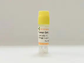
| Cat No. | ABC-TC0039 |
| Product Type | Human Gastric Cancer Cell Lines |
| Cell Type | Epithelial |
| Species | Human |
| Growth Conditions | 37 ℃, 5% CO2 |
| Source Organ | Stomach |
| Disease | Gastric Adenocarinoma |
| Product Code | AGS cell, AGS cell line |
The AGS cell line was derived from fragments of a tumor resected from a patient who had received no prior therapy.
AGS cell line is a human epithelial tumor cell line derived from gastric adenocarcinoma tissue obtained via surgical resection from a 54-year-old Caucasian female patient who had not received prior chemotherapy or radiotherapy. They exhibit a polygonal epithelial morphology and adhere tightly to culture surfaces in monolayer formation. They are characterized by a near-diploid karyotype with multiple structural and numerical chromosomal abnormalities associated with gastric carcinogenesis, including gains of chromosome 20q and losses of 18q. They retain functional p53 and exhibit moderate proliferation rates under standard culture conditions. AGS cells are tumorigenic but non-metastatic in immunodeficient mice and are widely used in gastric cancer studies. The cells undergo rigorous screening and isolation procedures, and are rigorously tested to ensure they are free of contamination from HIV-1, HBV, HCV, Syphilis, Mycoplasma, Fungi, Yeast, and Bacteria.
| Product Code | AGS cell, AGS cell line |
| Species | Human |
| Cat.No | ABC-TC0039 |
| Product Category | Tumor Cell Lines |
| Size/Quantity | 1 vial |
| Cell Type | Epithelial |
| Growth Mode | Adherent |
| Shipping Info | Dry Ice |
| Growth Conditions | 37 ℃, 5% CO2 |
| Source Organ | Stomach |
| Disease | Gastric Adenocarinoma |
| Biosafety Level | 2 |
| Storage | Liquid Nitrogen |
| Product Type | Human Gastric Cancer Cell Lines |
AGS cells are extensively utilized as a gastric adenocarcinoma model for studying oncogenic signaling, host-pathogen interaction, and epithelial transformation. Its epithelial origin and genetic stability make it ideal for research involving E-cadherin function, β-catenin signaling, and p53-related tumor suppression pathways. AGS is especially valuable in Helicobacter pylori pathogenesis studies. It is also employed in drug screening assays, cytotoxicity evaluation, and high-throughput screening for gastric cancer therapeutics. Due to its responsiveness to cytokines and growth factors, AGS provides a translational model for dissecting gastric epithelial biology, cancer development, and response to targeted treatments.
When you publish your research, please cite our product as “AcceGen Biotech Cat.# XXX-0000”. In return, we’ll give you a $200 coupon. Simply click here and submit your paper’s PubMed ID (PMID).
Maintaining the morphology of AGS cells involves several key steps to ensure they remain healthy and retain their characteristic appearance. Here’s how you can achieve this, along with methods to assess their health, viability, and perform morphological observations:
Maintaining Morphology of AGS Cells:
Culture Conditions: Maintain AGS cells in a suitable culture medium, typically DMEM (Dulbecco’s Modified Eagle Medium) supplemented with fetal bovine serum (FBS) and antibiotics (e.g., penicillin-streptomycin).
Subculture Regularly: Passage AGS cells when they reach about 70-80% confluence to prevent overgrowth, which can lead to changes in morphology. Follow recommended splitting ratios to avoid cell stress.
Monitor pH and Osmolarity: Ensure the pH of the medium is within the appropriate range (usually 7.2-7.4) and osmolarity is maintained, as fluctuations can affect cell morphology.
Avoid Over-Confluence: Cells should not be allowed to grow to full confluence where they become overly crowded, leading to differentiation or altered morphology.

Assessing Health and Viability of AGS Cells:
Trypan Blue Exclusion Assay: Mix AGS cells with Trypan Blue dye and count viable (unstained) versus non-viable (stained) cells under a microscope.
Cell Counting: Use automated cell counters or hemocytometers to count cells and assess changes in population density over time.
Metabolic Activity Assays: Perform assays such as MTT or MTS assays to measure mitochondrial activity, which correlates with cell viability.
Morphological Changes: Monitor cell morphology under a microscope regularly; healthy AGS cells typically appear epithelial with a polygonal shape and adherent.
Performing Morphological Observation of Cells:
Microscopy: Use an inverted phase-contrast microscope or an upright microscope with appropriate magnification (e.g., 10x to 40x objectives) to observe AGS cells.
Fixation and Staining: Fix cells with paraformaldehyde or methanol and stain with dyes like Hematoxylin and Eosin (H&E) for structural observation or fluorescent dyes for specific cellular components.
Live Cell Imaging: Use live-cell imaging techniques to observe dynamic changes in cell morphology and behavior over time without fixation.
Documentation: Record images or videos of AGS cells at different time points to track changes in morphology or response to treatments.