- In-Stock Tumor Cell Lines
- Human Orbital Fibroblasts
- Human Microglia
- Human Pulmonary Alveolar Epithelial Cells
- Human Colonic Fibroblasts
- Human Type II Alveolar Epithelial Cells
- Human Valvular Interstitial Cells
- Human Thyroid Epithelial Cells
- C57BL/6 Mouse Dermal Fibroblasts
- Human Alveolar Macrophages
- Human Dermal Fibroblasts, Adult
- Human Lung Fibroblasts, Adult
- Human Retinal Muller Cells
- Human Articular Chondrocytes
- Human Retinal Pigment Epithelial Cells
- Human Pancreatic Islets of Langerhans Cells
- Human Kidney Podocyte Cells
- Human Renal Proximal Tubule Cells
Respiration is a life-sustaining process in which gases are exchanged between the body and the outside atmosphere. Respiration is mainly carried out by the respiratory system.
The Respiratory System
Respiration by the respiratory system involves two subsidiary processes. One is breathing which is a physical process of conducting air to and from the lungs. The other process is gas exchange. This is a biochemical process in which oxygen diffuses out of the air and into the blood while carbon dioxide and other waste gases diffuse out of the blood and into the air. All organs of the respiratory system are involved in breathing, but only the lungs are involved in gas exchange [1][2].
The human respiratory system consists of a intricate network of tissues and organs, essential for the absorption of oxygen from the air and the elimination of carbon dioxide and other waste gases. Various cells within this system play pivotal roles, including maintaining epithelial homeostasis, preventing alveolar collapse, facilitating gas exchange between blood and tissues, and ensuring adaptation to low atmospheric pressure [3][4].
Primary cells obtained directly from human respiratory organs and tissues exhibit a remarkable ability to closely replicate in vivo conditions [5]. This fidelity enables researchers to gain profound insights into diverse lung diseases and associated medical disorders, significantly advancing our understanding of respiratory health.
Components of the respiratory system
The respiratory system is a physiological system composed of a series of organs and tissues. It is called the respiratory tract through which air flows into and out of the body. The respiratory tract has two major divisions: the upper respiratory tract and the lower respiratory tract. Figure 1 shows the main components of the respiratory system.
1.Upper respiratory tract: The upper respiratory tract consists of the nasal cavity, the pharynx, and the larynx. All these organsare involved in the conduction or the movement of air flow into and out of the body. They clean, humidify, and warm the incoming air and provide a route for air to move between the outside atmosphere and the lungs. There is no gas exchange occurs in these organs.
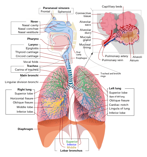
Figure 1. Components of the respiratory system [6].
2.Lower respiratory tract: The lower respiratory tract consists of the trachea, the primary bronchi and the lungs. Thetrachea and the bronchi in the lower respiratory tract conduct air between the upper respiratory tract and the lungs. Each bronchus branches into secondary bronchi, and so on. These passages form an inverted tree-like shape with repeated branching as moving deeper into the lungs (Figure 1). The lungs consist mainly of alveoli, which are the functional units of the lungs where gas exchange takes place. It is only in the lungs that gas exchange occurs between the air and the bloodstream [7, 8].
3.Besides these organs, certain muscles of the thorax are also involved inrespiration by enabling breathing. The most important one is the large muscle called the diaphragm, locating below the lungs and separating the thorax from the abdomen [9]. Contraction of the diaphragm increases the volume of the thoracic cavity, allowing air to be drawn into the lungs; relaxation of the diaphragm decreases the volume of the thoracic cavity, causing air to be expelled from the lungs.
Primary cell types of the respiratory system
The cells of the respiratory system cooperate to facilitate gas exchange, a vital life process taking place in alveoli, by transporting oxygen between the alveoli and the capillary network that surrounds them and delivering it to the body’s tissues, while simultaneously expelling carbon dioxide.
Primary cells of the respiratory system typically refer to cells isolated and purified from a wide variety of donors without the accumulated mutations found in immortal cell lines. Respiratory research benefits from these primary cells as they represent more physiological or pathological conditions. Here are some common types of primary cells in the respiratory system:
1.The respiratory epithelial cells: From the nasal cavity through the bronchi to the lung, therespiratory tract is covered in the epithelium. The airway epithelial cells play a unique role to external deleterious agents as a protective physical and functional barrier. Many diseases of the airway involve damage to the airway surface epithelium.
The bronchial epithelium is made up of the surface epithelial cells and mucus glands. The surface epithelial cells consist of three principle cell types: basal, goblet, and ciliated cells. These epithelial cells are crucial for studying infection, inflammation, and other diseases of the respiratory system.
Alveolar epithelial cells cover more than 99% of the internal surface of the lung and are also valuable cell models for study.
2.The vascular endothelial cells: These endothelial cellscontribute to the vascular homeostasis maintenance. They synthesize and secrete activators, inhibitors of both the fibrinolysis system and the coagulation system, and mediators which influence the blood platelets adhesion and aggregation. They also release molecules that can control cell proliferation and modulate vessel wall tone. The pulmonary microvascular endothelial cells function as a semiselective barrier that is critical for gas exchange and regulation of fluid and solute passage between the blood and interstitial compartments in the lungs.
3.Airway smooth muscle cells: Airway smooth muscle exists in the trachea and in the bronchial tree up to the terminal bronchioles. Their regulatory mechanisms and specialized features are the basis for normal airway function. In severe asthma patients, the characteristic feature is the increase in bronchial smooth muscle mass. Airway smooth muscle cells are also valuable models for studying diseases like bronchospasm, chronic bronchitis, and emphysema [10].
4.Fibroblasts: Fibroblasts,the most abundant cell type in lung interstitium, are one of the easiest types of cells to grow in culture. Pulmonary fibroblasts play an important role in the repair and remodeling processes following injury.
5.Pulmonarymacrophages: Pulmonary macrophages are widely distributed in the lung interstitium. They are active in phagocytosis, immunity and secretion and have important defence functions. Macrophages isolated from pulmonary interstitial tissues are essential components of the immune system responsible for clearing foreign particles and pathogens [11][12][13][14].
6.Epithelial stem cells: Stem cells present in the airway and lung epithelium, possessing the potential for self-renewal and differentiation into various epithelial cells [15][16].
Conclusion
The primary cells derived from the respiratory system are invaluable tools for numerous research applications. The utilization of them would significantly advance current research in uncovering the underlying mechanisms of various respiratory disorders and viral infections, offering critical insights into potential therapeutic avenues.
| Cat. No | Product Name | Cell Type | Price |
|---|---|---|---|
| ABC-TC5515 | Human Type II Alveolar Epithelial Cells | Epithelial Cells | +Inquiry |
| ABC-TC3770 | Human Pulmonary Alveolar Epithelial Cells | Epithelial Cells | +Inquiry |
| ABC-TC3774 | Human Pulmonary Artery Endothelial Cells | The Vascular Endothelial Cells | +Inquiry |
| ABC-TC3530 | Human Bronchial Smooth Muscle Cells | Airway Smooth Muscle Cells | +Inquiry |
| ABC-TC3683 | Human Lung Fibroblasts, Adult | Fibroblasts | +Inquiry |
| ABC-H0034X | Human Alveolar Macrophages | Pulmonary Macrophages | +Inquiry |
References
[1] D.W. Empey, Diseases of the respiratory system. Introduction: structure and function of the lungs, Br Med J 1(6113) (1978) 631-3.
[2] M. Bastir, D. Sanz-Prieto, J.M. Lopez-Rey, C.A. Palancar, M. Gomez-Recio, M. Lopez-Cano, J.M. Gonzalez-Ruiz, A. Perez-Ramos, M.A. Burgos, B. Beyer, D. Garcia-Martinez, The evolution, form and function of the human respiratory system, J Anthropol Sci 100 (2022) 141-172.
[3] D. Siegler, The respiratory system. 1. Function and structure, Nurs Mirror 145(24) (1977) 13-4.
[4] [The defense mechanism of the respiratory system and its control], Nihon Kyobu Shikkan Gakkai Zasshi 19(12) (1981) 925-63.
[5] A. Masui, T. Hirai, S. Gotoh, Perspectives of future lung toxicology studies using human pluripotent stem cells, Arch Toxicol 96(2) (2022) 389-402.
[6] J. Sobotta, Atlas der deskriptiven Anatomie des Menschen, 10. Aufl. ed., Urban & Schwarzenberg, Berlin, 1946.
[7] K. Tetzlaff, Pulmonary Physiology and Medicine of Diving, Semin Respir Crit Care Med 44(5) (2023) 705-718.
[8] D.W. Kaczka, J.H. Bates, Pulmonary physiology. Preface, Crit Rev Biomed Eng 39(4) (2011) 261-2.
[9] M. Gama de Abreu, F.J. Belda, Neurally adjusted ventilatory assist: letting the respiratory center take over control of ventilation, Intensive Care Med 39(8) (2013) 1481-3.
[10] R.A. Panettieri, Jr., M.I. Kotlikoff, W.T. Gerthoffer, M.B. Hershenson, P.G. Woodruff, I.P. Hall, S. Banks-Schlegel, L. National Heart, I. Blood, Airway smooth muscle in bronchial tone, inflammation, and remodeling: basic knowledge to clinical relevance, Am J Respir Crit Care Med 177(3) (2008) 248-52.
[11] N. Joshi, J.M. Walter, A.V. Misharin, Alveolar Macrophages, Cell Immunol 330 (2018) 86-90.
[12] T.A. Poor, L. Morales-Nebreda, Alveolar Macrophages during Inflammation: A Balancing Act, Am J Respir Cell Mol Biol 68(6) (2023) 608-609.
[13] C.Y. Chang, D. Armstrong, D.B. Corry, F. Kheradmand, Alveolar macrophages in lung cancer: opportunities challenges, Front Immunol 14 (2023) 1268939.
[14] F.P. Martin, C. Jacqueline, J. Poschmann, A. Roquilly, Alveolar Macrophages: Adaptation to Their Anatomic Niche during and after Inflammation, Cells 10(10) (2021).
[15] S.H. Randell, Airway epithelial stem cells and the pathophysiology of chronic obstructive pulmonary disease, Proc Am Thorac Soc 3(8) (2006) 718-25.
[16] Q. Chen, Y. Liu, Isolation and culture of mouse alveolar type II cells to study type II to type I cell differentiation, STAR Protoc 2(1) (2021) 100241.
Copyright - Unless otherwise stated all contents of this website are AcceGen™ All Rights Reserved – Full details of the use of materials on this site please refer to AcceGen Editorial Policy – Guest Posts are welcome, by submitting a guest post to AcceGen you are agree to the AcceGen Guest Post Agreement – Any concerns please contact marketing@accegen.com