




For research use only
| Cat No. | ABC-TC0640 |
| Product Type | Human Breast Cancer Cell Lines |
| Species | Human |
| Growth Conditions | 37 ℃, 5% CO2 |
| Product Code | MCF 7, MCF-7, MCF.7, MCF7 cell, MCF-7 cell, MCF 7 cell |
MCF7 Breast Cancer Cells are ER-positive and used for hormone-driven breast cancer modeling, WNT signaling, and epithelial behavior studies.
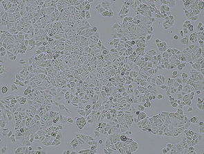
MCF-7 is a human breast cancer cell line derived from the pleural effusion of a 69-year-old Caucasian female with metastatic breast adenocarcinoma. The cells are estrogen receptor-positive (ER⁺), progesterone receptor-positive (PR⁺), and HER2-negative, representing the luminal A subtype of breast cancer. They are slow-growing, form loosely attached three-dimensional clusters with floating viable cells, and may show delayed attachment after initial passaging. Growth is inhibited by tumor necrosis factor alpha (TNF-α). Cytogenetically, they exhibit a near-diploid karyotype (46–50 chromosomes) and can form tumors in estrogen-supplemented nude mice. These cells exhibit features of differentiated mammary epithelium, including the ability to respond to estradiol via nuclear estrogen receptor α (ERα) and form epithelial domes. They also express epithelial markers including E-cadherin and cytokeratins. MCF-7 cells overexpress WNT7B (not classified as an oncogene), insulin-like growth factor binding proteins IGFBP-2, -4, and -5, and intracellular adhesion molecule 1 (ICAM-1), which contributes to colony formation and cell adhesion through NF-κB regulation.
| Product Code | MCF 7, MCF-7, MCF.7, MCF7 cell, MCF-7 cell, MCF 7 cell |
| Species | Human |
| Cat.No | ABC-TC0640 |
| Product Category | Tumor Cell Lines |
| Size/Quantity | 1 vial |
| Shipping Info | Dry Ice |
| Growth Conditions | 37 ℃, 5% CO2 |
| Biosafety Level | 1 |
| Storage | Liquid Nitrogen |
| Product Type | Human Breast Cancer Cell Lines |
MCF-7 is a widely used estrogen receptor-positive (ER⁺) breast cancer cell line instrumental in studying hormone signaling and therapeutic response. It expresses functional progesterone receptors (PR) and shows variable androgen (AR) and glucocorticoid receptor (GR) expression across passages. This enables exploration of multiple steroid pathways relevant to breast cancer progression and resistance to ER-targeted therapies.
When you publish your research, please cite our product as “AcceGen Biotech Cat.# XXX-0000”. In return, we’ll give you a $200 coupon. Simply click here and submit your paper’s PubMed ID (PMID).
Oh CS, Hong SW, Park S, Kwon Y, Kim SH. Effect of equipotent doses of propofol and sevoflurane on endoplasmic reticulum stress during breast cancer surgery. Korean J Anesthesiol. 2022;75(6):487-495. doi:10.4097/kja.21569
1. Preparation
Warm the DMEM or RPMI-1640 medium, PBS, and trypsin-EDTA solution to 37°C. Ensure all reagents and equipment are sterile and ready for use.
2. Observation
Place the flask containing MCF7 cells under an inverted microscope. Check for cell confluence (typically, cells should be 70-80% confluent before passaging).
3. Removing Old Medium
In a biosafety cabinet, carefully aspirate the old medium without disturbing the cell monolayer.
4. Rinsing Cells
Add 5-10mL of PBS to the flask to rinse off any residual medium and serum that may inhibit trypsin activity. Gently rock the flask to ensure the cells are thoroughly rinsed, then aspirate the PBS.
5. Adding Trypsin-EDTA
Add enough trypsin-EDTA solution to cover the cell monolayer (typically 1-2mL for a T-75 flask). Gently tilt the flask to ensure the entire surface is covered with trypsin-EDTA.
6. Incubation
Place the flask in the incubator for 3-5 minutes, checking the cells under the microscope periodically. The cells should round up and detach from the surface. Avoid over-trypsinization, which can damage the cells and affect their differentiation state.
7. Neutralizing Trypsin
Once the cells are detached, add 5-10 mL of complete medium (with FBS) to the flask to neutralize the trypsin.
8. Collecting Cells
Gently pipette the cell suspension up and down to ensure all cells are detached and in suspension. Transfer the cell suspension to a 15mL conical tube.
9. Centrifugation
Centrifuge the cell suspension at 200×g for 5 minutes to pellet the cells.
10. Resuspending Cells
Carefully aspirate the supernatant without disturbing the cell pellet. Resuspend the cells in an appropriate volume of fresh complete medium.
11. Seeding New Flasks
Seed the cells into new culture flasks or plates at the desired density. For example, if splitting at a 1:5 ratio, add 1 part of the cell suspension to 4 parts of fresh medium in a new flask. Avoid seeding at too high or too low a density, as this can affect cell growth and differentiation.
12. Incubation
Place the newly seeded flasks or plates back into the incubator set at 37°C with 5% CO2.
13. Monitoring
Observe the cells under the microscope after 24 hours to ensure they have adhered and are spreading out. Change the medium every 2-3 days or as needed.
Tips for Maintaining Differentiation State
Serum Quality: Use high-quality, consistent batches of FBS, as variations can affect cell differentiation.
Gentle Handling: Handle cells gently during trypsinization and resuspension to minimize stress and avoid differentiation.
Consistent Conditions: Maintain consistent culture conditions (temperature, CO2 levels, and medium composition) to prevent stress-induced differentiation changes.
Minimal Passages: Keep the passage number low, as high passage numbers can lead to genetic drift and changes in cell behavior, including differentiation.
Avoid Confluence: Do not allow cells to become overly confluent, as this can lead to differentiation and changes in cell behavior.
To assess the differentiation status of MCF7 cells, a widely used human breast cancer cell line, several key markers can be evaluated. These markers include proteins, receptors, and genes associated with specific cellular functions and phenotypes that indicate differentiation. Here are the key markers and methods to assess the differentiation status of MCF7 cells:
Key Markers
1. Estrogen Receptor (ER)
Description: Estrogen receptor alpha (ERα) is a key marker for the luminal subtype of breast cancer, to which MCF7 cells belong.
Assessment: ERα expression can be evaluated using immunohistochemistry (IHC), Western blotting, or quantitative PCR (qPCR).
2. Progesterone Receptor (PR)
Description: PR is another important hormone receptor expressed in luminal breast cancer cells, including MCF7 cells.
Assessment: Similar to ER, PR expression can be assessed using IHC, Western blotting, or qPCR.
3. E-Cadherin
Description: E-cadherin is a cell adhesion molecule important for maintaining epithelial cell phenotype and preventing epithelial-mesenchymal transition (EMT).
Assessment: E-cadherin expression is typically measured by IHC, Western blotting, or qPCR.
4. Cytokeratins (CKs)
Description: Cytokeratins such as CK8 and CK18 are intermediate filament proteins expressed in epithelial cells.
Assessment: Cytokeratin expression can be assessed using IHC, Western blotting, or qPCR.
5. GATA3
Description: GATA3 is a transcription factor involved in maintaining the differentiation of luminal epithelial cells.
Assessment: GATA3 expression can be evaluated using IHC, Western blotting, or qPCR.
For culturing MCF7 cells, a commonly used human breast cancer cell line, the recommended culture medium is Dulbecco’s Modified Eagle Medium (DMEM) supplemented with appropriate additives to support optimal cell growth and maintenance.