- In-Stock Tumor Cell Lines
- Human Orbital Fibroblasts
- Human Microglia
- Human Pulmonary Alveolar Epithelial Cells
- Human Colonic Fibroblasts
- Human Type II Alveolar Epithelial Cells
- Human Valvular Interstitial Cells
- Human Thyroid Epithelial Cells
- C57BL/6 Mouse Dermal Fibroblasts
- Human Alveolar Macrophages
- Human Dermal Fibroblasts, Adult
- Human Lung Fibroblasts, Adult
- Human Retinal Muller Cells
- Human Articular Chondrocytes
- Human Retinal Pigment Epithelial Cells
- Human Pancreatic Islets of Langerhans Cells
- Human Kidney Podocyte Cells
- Human Renal Proximal Tubule Cells
What is Tonsil?
Tonsil are a group of lymphoid located into the aerodigestive tract, also called Waldeyer’s tonsillar ring[1]. There are 4 types of tonsils for human, including the pharyngeal tonsil, two tubal tonsils, two palatine tonsils and the lingual tonsils[2]. Tonsils are important immunocompetent organs and are identified as the first line of defense for the infection of exogenous pathogens[3]. M cells (specialized antigen capture cells) exist on the surface of tonsils that can capture antigen and stimulate the immune response through B cells and T cells[1].
Common pathological changes of tonsils include inflammation (tonsillitis) or enlarged palatine tonsils (adenotonsillar hyperplasia, mainly caused by clinical inflammation or inflammation recurring), and tumors or cancer are also possible[1]. Therapy method of inflammation of tonsil mainly symptomatic drug therapy, surgical removal may be considered if the tonsil blocks the airway or repeated bouts of inflammation[4].

Figure1. Tonsils
Introduction and Culture Protocol of Human Tonsil Cells
Human Tonsil Primary Lymphatic Microvascular Endothelial Cells
The lymphatic system is a complex network of numerous microvessels and throughout most organs of the whole body[5]. Lymphatic microvascular endothelial cells (LMEC) can be isolated from various human organs or tissue such as derma, lymphatic vascular tumors, and palatine tonsils[6-8]. And palatine tonsil become a better choice for the isolation of LMEC thanks to the easy surgical access of palatine tonsil tissue. Besides, the growth of LMEC is selectively regulated by VEGF-C through VEGFR-3[8].
The isolation protocol of human primary LMEC will be briefly described here[8]. After trypsinization, the primary culture is collected and centrifugated and then treated by magnetic beads with anti-CD31 mAb or magnetic tosyl-activated beads coated with lectin UEA-1 to enrich for LMEC. After magnetic beads purification, the cells were culture in an endothelial growth medium with VEGF-C. Human primary LMEC isolated from human palatine tonsils can be cultured in vitro 8-10 generations, and the recommended split ratio for each passage is 1:3. Besides, human primary LMEC recommended for experiments are kept below passage 5.
Human Tonsil Epithelial Cells
Tonsil epithelium is a heterogeneous lymphoid organ and can format surface squamous epithelium and crypts. The crypts of the tonsil are a special structure that plays role in the immune response of the tonsil[9]. Besides, the crypts are also easy to accumulate tonsil stone, which is consists of food scraps, bacteria, or mucosal secretions. And the accumulation of tonsil stone is also one of the inflammation stimuli of tonsil diseases.
About the isolation and culture of the human tonsil epithelial cells (HTEpiC), here is a brief introduction to the protocol by taking human primary tonsil epithelial keratinocytes as an example[10]. After tissue separation, the mucosal epithelial layers are treated with collagenase and dispase to get epithelial cells. After being washed by the medium 2 times, the cells are dissociated into single cells in trypsin/EDTA, and then the culture is filtered to remove debris. Finally, after the cells are washed by the culture medium then let cells grow in the medium.
Applications of Primary Human Tonsil Cells
Human Tonsil Primary Lymphatic Microvascular Endothelial Cells
Lymphatic microvascular endothelial cells isolated from the tonsil are a valuable model for the research of lymphatic vessels. Caruso, et al. use human tonsil primary lymphatic microvascular endothelial cells (LECs) to identify the inhibition of the infection of human herpesvirus 6 (HHV-6) to the lymphangiogenesis and angiogenesis[11]. They isolate and culture human primary LECs in vitro[8] and infect the cells with HHV-6, and find that HHV-6 infection inhibits the formation of capillary-like structures. Further, they identify the expression of U94/rep coding by HHV-6 as the culprit leading to the disorder of vascular structure generation. Besides, they identify U94/rep U94/rep and its gene product affect LECs migration, and the conclusion of this research is validated with a rat model.
Human Tonsil Epithelial Cells
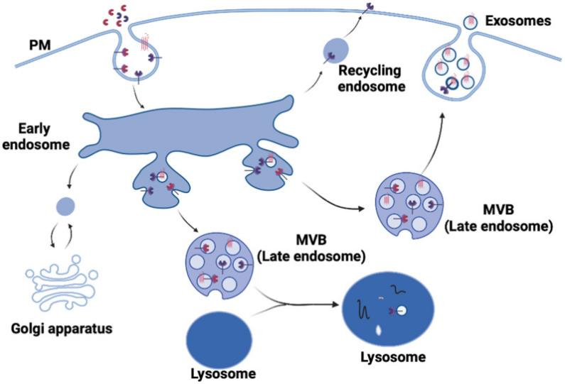
The mechanism of mother-to-child transmission (MTCT) of HIV-1 is a complex pathological process, and tonsil is an important link in this process. Herrera, et al. use human primary tonsil epithelial cells (hTECs) to identify the beta-defensins 2 and 3 (hBD-2, hBD-3) that can inactivate HIV-1 in infant tonsil epithelial cells[10]. To let hBD inactivate HIV-1 in the multivesicular bodies (MVB) and vacuoles in hTECs, they tagged hBDs with the protein transduction domain (PTD) of HIV-1 Tat to let hBDs easier to be transported to where HIV is located. They found that hBD-2 and hBD-3 labeled by PTD can efficiently penetrate cellular barriers and internalization into MVB and vacuoles containing HIV-1 in hTECs, and PTD plays a key role in the process of the fusion of vesicles containing HIV-1 with lysosomes, leading to the inactivation of HIV-1 in hTECs. Besides, they use ex vivo tonsil tissue explants to identify that hBD labeled by PTD decreases the spread of HIV-1 from the tonsil to the CD4+ T lymphocytes, CD68+ macrophages, and CD1c+ dendritic cells. All in all, they use hTECs to find hBD-2 and hBD-3 labeled by PTD is a new strategy in reducing viral MTCT of HIV-1.
Conclusion
As for the first line of defense of the immune system in the aerodigestive tract, the tonsil plays a key role in defending exogenous pathogens. And since tonsillectomy is a widely used treatment in clinical practice, the tonsil is one of the most accessible models of lymphoid organs. Therefore, various kinds of human primary tonsil cells become an ideal model for immune system research.
Where to Get Primary Human Tonsil Cells for research?
Currently, AcceGen provides 3 types of Human Tonsil Cells for research purposes.
| CAT.# | NAME |
| ABC-TC3834 | Human Tonsil Epithelial Cells |
| ABC-TC3835 | Human Tonsil Fibroblasts |
| ABC-TC3836 | Human Tonsil Microvascular Endothelial Cells |
To get more information, please refer to: Tonsil Cells.
It is our pleasure to help relative researches to move forward. All the products of AcceGen are strictly comply with international standards. For more detailed information, please visit our product portfolio or contact [email protected].
Reference
1. Kato A, Hulse KE, Tan BK, Schleimer RP: B-lymphocyte lineage cells and the respiratory system.J Allergy Clin Immunol 2013, 131:933-957; quiz 958.
2. Universities of Fribourg LaBS: Tonsil.https://embryology.ch/en/organogenesis/digestion-tract/face-and-upper-foregut/definitive-pharynx/tonsils.html?p=6.2#tonsils.
3. Scadding GK: Immunology of the tonsil: a review.J R Soc Med 1990, 83:104-107.
4. MedlinePlus: Adenoids.https://medlineplus.gov/adenoidshtml.
5. Ryan TJ, Curri SB: Blood vessels and lymphatics.Clin Dermatol 1989, 7:25-36.
6. Kriehuber E, Breiteneder-Geleff S, Groeger M, Soleiman A, Schoppmann SF, Stingl G, Kerjaschki D, Maurer D: Isolation and characterization of dermal lymphatic and blood endothelial cells reveal stable and functionally specialized cell lineages.J Exp Med 2001, 194:797-808.
7. Mancardi S, Stanta G, Dusetti N, Bestagno M, Jussila L, Zweyer M, Lunazzi G, Dumont D, Alitalo K, Burrone OR: Lymphatic endothelial tumors induced by intraperitoneal injection of incomplete Freund’s adjuvant.Exp Cell Res 1999, 246:368-375.
8. Garrafa E, Alessandri G, Benetti A, Turetta D, Corradi A, Cantoni AM, Cervi E, Bonardelli S, Parati E, Giulini SM, et al: Isolation and characterization of lymphatic microvascular endothelial cells from human tonsils.J Cell Physiol 2006, 207:107-113.
9. Roberts S, Evans D, Mehanna H, Parish JL: Modelling human papillomavirus biology in oropharyngeal keratinocytes.Philos Trans R Soc Lond B Biol Sci 2019, 374:20180289.
10. Herrera R, Rosbe K, Tugizov SM: Inactivation of HIV-1 in Polarized Infant Tonsil Epithelial Cells by Human Beta-Defensins 2 and 3 Tagged with the Protein Transduction Domain of HIV-1 Tat.Viruses 2021, 13.
11. Caruso A, Caselli E, Fiorentini S, Rotola A, Prandini A, Garrafa E, Saba E, Alessandri G, Cassai E, Di Luca D: U94 of human herpesvirus 6 inhibits in vitro angiogenesis and lymphangiogenesis.Proc Natl Acad Sci U S A 2009, 106:20446-20451.
Copyright - Unless otherwise stated all contents of this website are AcceGen™ All Rights Reserved – Full details of the use of materials on this site please refer to AcceGen Editorial Policy – Guest Posts are welcome, by submitting a guest post to AcceGen you are agree to the AcceGen Guest Post Agreement – Any concerns please contact [email protected]