Home > Recent Posts >

<<Previous Next>>

Post Date:Nov-19-19

Skeletal Muscle Satellite Cells and Macrophages Regulate Skeletal Muscle Regeneration

AcceGenAuthor: AcceGen R&D Team

Abstract

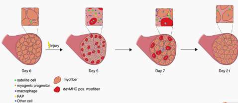
Skeletal Muscles are composed of many contractile muscle cells and are covered by connective tissue. During embryonic and fetal development, muscle stem cells fuse to form muscle fibers. Skeletal Muscle Cells are composed of Skeletal Muscle Stem Cells, Skeletal Muscle Satellite Cells (Skeletal Muscle SCs) , Fibro/Adipogenic Progenitors (FAPs), Macrophages, Neutrophils, Endothelial Cells, C2C12 Skeletal Muscle Myoblast and so on[1]. The functions of different cells in skeletal muscle regeneration were researched.

Product Background

Skeletal muscle is the most important organ of human beings playing an important role in physical activity[2]. Skeletal muscle has a remarkable capacity to regenerate even after repeated traumas, yet limited information is available on muscle repair mechanisms and how they have evolved.

Skeletal regeneration is a very complex biological process in which skeletal muscle stem cells participate in the repair process[3]. Skeletal muscle repair and regeneration rely on driving muscle development to help muscle regeneration.

Muscle Stem Cells (Satellite Cells), Mononuclear Progenitor Cells and Macrophages are the basis of skeletal muscle tissue. Therefore, the studies of skeletal muscle cell types and their respective functions in skeletal muscle regeneration make a major contribution in the treatment of bone diseases.

The Latest Research

  • 1. Satellite Cells are Considered the Primary Player of Regenerative Myogenesis, Adult Skeletal Muscle Maintains a Relatively Remarkable Ability to Regenerate and Recover Following Mild Damage or Insult throughout Much of the Lifespan.


    Application: Skeletal muscle satellite cells do not cause muscle atrophy that suggested its primary function in adults is bone regeneration. The satellite community is considered to be responsible for post-natal muscle growth and tissue regeneration after appropriate stimulation. The research shows TF Yin Yang1 and N-methyltransferase inhibitors can regulate skeletal muscle regeneration through controlling the metabolic reprogramming of satellite cells.

Satellite cells were first discovered by electron microscopy in 1961 by Alexander Mauro on the periphery of skeletal muscle myofibers [4]. Satellite cells are derived from specific locations within muscle tissue that are closely related to individual muscle fibers.

Finnerty and colleagues reported that the recovery of skeletal muscle burns depends on the activity of satellite cells while the conditional consumption of satellite cells reduces the regeneration of atrophic muscles[5].

Chen et al. found that YY1 was identified as a key regulator of SC metabolic reprogramming by regulating the dual roles of mitochondria and glycolytic pathways. SC examination showed that loss of YY1 resulted in autonomous proliferation defects in activated cells that indicated increasing YY1 expression promotes skeletal muscle regeneration[6].

Harshini et al. discovered that inhibition of small molecule nicotinamide N-methyltransferase inhibitors enhances muscle fiber regeneration and growth after injury and activates senescent muscle stem cells, improving skeletal muscle regeneration in the elderly[7].

Lee et al. showed the role of autophagy in skeletal muscle regeneration while emphasizing the revitalization strategy of altering autophagy to improve muscle regeneration[8].

An interesting study discovered that elderly rodent skeletal muscle fibers were transplanted into the muscles of young rats which the satellite cells remained intact and restored the ability of the aged satellite cells to regenerate[9].


  • During muscle regeneration, the time course of changes in cellular composition during skeletal muscle regeneration following cardiotoxin (CTX) injury.


    Figure1. During muscle regeneration, the time course of changes in cellular composition during skeletal muscle regeneration following cardiotoxin (CTX) injury. Satellite cells (in green) are quiescent in resting skeletal muscle. Five days after CTX injury, regenerating muscles are reduced to mostly mononuclear cells (satellite cells immune cells, etc.), but are able to form new myotubes at day 7, which mature to multinucleated myofbers[2].

  • 2. Skeletal Muscle Macrophages are Closely Related to The Pro-inflammatory and Recovery Stages of Tissue Repair in the Body and Can be Recruited into Damaged Areas in the Vascular Phase.

    Application: Macrophages can make bones vital to muscle regeneration and promote the coordination of many bioremediation processes. The hepatocyte growth factor can through CaMKKβ-AMPK signaling promote skeletal muscle regeneration.

Skeletal muscle macrophages are complex populations of immune cells throughout the body. Macrophages are usually classified into pro-inflammatory (M1) or anti-inflammatory (M2) depending on the expression of surface markers[10]. macrophages are key regulators of different biological processes involved during skeletal muscle regeneration, such as myogenesis, fibrosis, inflammation, and revascularization[11].

Walton et al. found that skeletal muscle macrophages did not respond to RE, and in the absence of severe tissue damage that intramuscular macrophages were more polarized in the M2 direction than previously recognized[12].

Choi et al. demonstrated that the HGF/c-met pathway plays a key role in skeletal muscle macrophages during muscle regeneration following necrotic injury. The macrophage was the major cell type affected by HGF among cells that infiltrated the muscle and HGF/c-met signaling is the principle part in the transition of the macrophage infiltrated during muscle regeneration[13].

Sorensen et al. studied macrophage content and phenotype following exercise-induced muscle injury regimens in young and elderly patients. At the same time, they also used primary cell culture models to examine the effects of macrophage secretion and juvenile macrophage conditioned medium on proliferation and differentiation potential[10].

Minar et al. evaluated two classical mouse macrophage subtypes and some post-exercise inflammatory cytokine findings associated with muscle adaptation processes. Increased Ly6C + macrophages and IL-6 and IL-13 may help us understand the mechanism between inflammation and regeneration [14].

co-expression of both M1 and M2 macrophage markers


Figure2. Flow cytometry from discarded human hamstring muscle showing co-expression of both M1 and M2 macrophage markers. Mononuclear cells isolated from human skeletal muscle express A. Both pan-monocyte markers CD11b and CD14; B. Both the M2 macrophage marker, CD206, and the pan marker, CD11b; C. Both the M1 marker, CD86, and the pan marker CD11b; D. Both the M2 marker, CD206, and the M1 marker, CD86; E. Overlay of CD206+/CD86+ populations from panel D onto CD14/CD11b flow plot shown in panel A. This overlay shows that CD206+/CD86+ macrophages (denoted in dark blue) also express the pan-monocyte markers CD11b and CD14. Red boxes indicate cells that are double positive for the markers shown[15].

Conclusion

Skeletal muscle regeneration is a highly coordinated process that activates multiple factors and multiple cell types to remove necrotic cell debris and repair muscle fiber damage. The satellite and other functional cell types play important roles in muscle repair and regeneration. Several types of cells, including SCs, are directly involved in generating new myofibers, while other cell types secrete factors that regulate SC fate and regeneration.
Furthermore, the interaction and crosstalk among the different cell types during the regenerative process have not been fully elucidated. Therefore, future studies are needed to obtain a more comprehensive understanding of the skeletal muscle regenerative process, as well as provide a basis for the development of better therapies for muscular disorders.

AcceGen Skeletal Muscle Primary Cells

AcceGen’s primary cell repository contains Skeletal Muscle Cells with various species, various cell types, and various growth stages of donors.

Choose the one that meets your specific research needs or contact us at 1-862-686-2696 or inquiry@accegen.com for more detailed information:


Human Skeletal Muscle Cells: Normal Postnatal Prenatal Uncultured Foetal Pre-screened Foetal
Human Skeletal Muscle Satellite CellsHuman Skeletal Muscle Progenitor CellsHuman Skeletal Muscle MyoblastsHuman Skeletal Myoblasts


Rat Skeletal Muscle Cells: Adult Foetal Canine Skeletal Muscle CellsFeline Skeletal Muscle Cells


Reference

1. Chen B, Shan T: The role of satellite and other functional cell types in muscle repair and regeneration. J Muscle Res Cell Motil 2019, 40(1):1-8.

2. Schmidt M, Schuler SC, Huttner SS, von Eyss B, von Maltzahn J: Adult stem cells at work: regenerating skeletal muscle. Cell Mol Life Sci 2019, 76(13):2559-2570.

3. Cho DS, Doles JD: Skeletal Muscle Progenitor Cell Heterogeneity. Adv Exp Med Biol 2019, 1169:179-193.

4. A M: . J Biophys Biochem Cytol 1961.

5. Finnerty CC, McKenna CF, Cambias LA, Brightwell CR, Prasai A, Wang Y, El Ayadi A, Herndon DN, Suman OE, Fry CS: Inducible satellite cell depletion attenuates skeletal muscle regrowth following a scald-burn injury. J Physiol 2017, 595(21):6687-6701.

6. Chen F, Zhou J, Li Y, Zhao Y, Yuan J, Cao Y, Wang L, Zhang Z, Zhang B, Wang CC et al: YY1 regulates skeletal muscle regeneration through controlling metabolic reprogramming of satellite cells. EMBO J 2019, 38(10).

7. Neelakantan H, Brightwell CR, Graber TG, Maroto R, Wang HL, McHardy SF, Papaconstantinou J, Fry CS, Watowich SJ: Small molecule nicotinamide N-methyltransferase inhibitor activates senescent muscle stem cells and improves regenerative capacity of aged skeletal muscle. Biochem Pharmacol 2019, 163:481-492.

8. Lee DE, Bareja A, Bartlett DB, White JP: Autophagy as a Therapeutic Target to Enhance Aged Muscle Regeneration. Cells 2019, 8(2).

9. John K. Hall GBB, 2 Jeffrey S. Chamberlain,2 Bradley B. Olwin: Prevention of Muscle Aging by Myofiber-Associated Satellite Cell Transplantation. science 2010, Vol 2 Issue 57 57ra83.

10. Sorensen JR, Kaluhiokalani JP, Hafen PS, Deyhle MR, Parcell AC, Hyldahl RD: An altered response in macrophage phenotype following damage in aged human skeletal muscle: implications for skeletal muscle repair. FASEB J 2019, 33(9):10353-10368.

11. Dort J, Fabre P, Molina T, Dumont NA: Macrophages Are Key Regulators of Stem Cells during Skeletal Muscle Regeneration and Diseases. Stem Cells Int 2019, 2019:4761427.

12. Walton RG, Kosmac K, Mula J, Fry CS, Peck BD, Groshong JS, Finlin BS, Zhu B, Kern PA, Peterson CA: Human skeletal muscle macrophages increase following cycle training and are associated with adaptations that may facilitate growth. Sci Rep 2019, 9(1):969.

13. Choi W, Lee J, Lee J, Lee SH, Kim S: Hepatocyte Growth Factor Regulates Macrophage Transition to the M2 Phenotype and Promotes Murine Skeletal Muscle Regeneration. Front Physiol 2019, 10:914.

14. André Luis Araujo Minari1 FA, Lila Missae Oyama3, Ronaldo Vagner Thomatieli-Santos2.: Skeletal muscle induce recruitment of Ly6C+ macrophages subtype and release inflammatory cytokines 3 days after downhill exercise. physiology 2019.

15. Kosmac K, Peck BD, Walton RG, Mula J, Kern PA, Bamman MM, Dennis RA, Jacobs CA, Lattermann C, Johnson DL et al: Immunohistochemical Identification of Human Skeletal Muscle Macrophages. Bio Protoc 2018, 8(12).

Leave a Reply

Your email address will not be published.

<<Previous Next>>

AcceGen Scroll Top Button
Copy link