



PD-1/PD-L1 Checkpoint Blockade
Evaluate PD-1/PD-L1 pathway inhibition and restoration of immune-mediated tumor killing using functional reporter and tumor cell models.
Evaluate immune checkpoint blockade and checkpoint-dependent tumor killing using ready-to-use tumor cell models and immune cell systems for immunotherapy research.
Explore Promotion Products →
Evaluate immune checkpoint-dependent tumor killing using cell-based models supporting checkpoint inhibition, immune reactivation, cytotoxicity, and therapeutic response evaluation.
Evaluate PD-1/PD-L1 pathway inhibition and restoration of immune-mediated tumor killing using functional reporter and tumor cell models.
Characterize CTLA-4-mediated immune suppression and functional recovery following checkpoint blockade.
Measure tumor cell killing following checkpoint inhibition using functional cytotoxicity and reporter-based assays.
Compare checkpoint inhibitor activity and downstream immune responses across different therapeutic candidates.
Checkpoint-dependent tumor killing involves immune suppression, checkpoint blockade, immune reactivation, and tumor cell elimination. AcceGen provides complementary cell models to support key stages of this workflow.
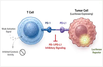
PD-1 on T cells engages PD-L1 on tumor cells, suppressing T cell activation and downstream signaling.
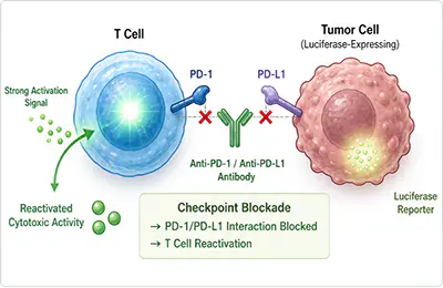
Anti-PD-1 or anti-PD-L1 antibodies disrupt receptor–ligand interactions, restoring immune signaling and T cell effector activity.
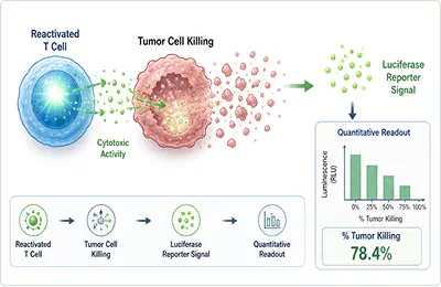
Restored immune activity leads to tumor cell elimination, which can be quantitatively assessed using luciferase reporter models and other functional readouts.
Beyond PD-1/PD-L1, AcceGen provides models supporting CTLA-4, LAG-3, and TIGIT checkpoint research.
Explore complementary cell models supporting immune checkpoint signaling, immune reactivation, tumor killing, and quantitative cytotoxicity assessment.
Wild-type and luciferase-expressing tumor cells for checkpoint-dependent killing studies and quantitative assessment of tumor cell killing.
Functional reporter cells for evaluating PD-1/PD-L1, CTLA-4, LAG-3, TIGIT, T cell activation, and related immune signaling pathways.
PBMCs, purified T cells, and other immune cell models for functional checkpoint blockade and immune-mediated cytotoxicity studies.

Explore selected cell lines for T cell-mediated killing, CAR-T, and immunotherapy research.
| CAT.# | Product / Cell Line | Cell Origin | Key Features | Recommended Applications | Lead Time |
|---|---|---|---|---|---|
| ABC-TC621S | MC38 | Murine colorectal carcinoma | PD-L1-positive | PD-1/PD-L1 blockade evaluation | 1–3 weeks |
| ABC-TC0197 | CT-26 | Murine colorectal carcinoma | PD-L1-positive | PD-1/PD-L1 blockade evaluation | 1–3 weeks |
| ABC-TC0062 | B16-F10 | Murine melanoma | Low basal PD-L1 expression; inducible upon stimulation | Immune checkpoint research model | 1–3 weeks |
| ABC-TC0026 | A375 | Human melanoma | PD-L1-positive | PD-1/PD-L1 blockade assays | 3–5 weeks |
| ABC-TC0032 | A549 | Human non-small cell lung carcinoma | PD-L1-positive | PD-1/PD-L1 blockade assays | 1–3 weeks |
| ABC-TC0337 | HCT116 | Human colorectal carcinoma | PD-L1-positive | Immune checkpoint research model | 1–3 weeks |
| ABC-TC0924 | Raji | Human Burkitt lymphoma | PD-L1-positive; inducible upon stimulation | Immune checkpoint co-culture assays | 1–3 weeks |
| ABC-TC1258 | U-87 MG | Human glioblastoma | PD-L1-positive | Neuro-oncology immune checkpoint studies | 1–3 weeks |
| ABC-TC0651 | MDA-MB-231 | Human triple-negative breast cancer | PD-L1-positive | Breast cancer immune checkpoint studies | 1–3 weeks |
| ABC-TC0591 | LLC | Murine lung carcinoma | PD-L1-positive | Murine lung cancer model studies | 1–3 weeks |
| ABC-RC193H | MC38-Luc | Murine colorectal carcinoma | Constitutive luciferase reporter cell line | PD-1/PD-L1 blockade evaluation | In stock |
| ABC-RC587H | CT-26-Luc | Murine colorectal carcinoma | Constitutive luciferase reporter cell line | PD-1/PD-L1 blockade evaluation | 2–3 weeks |
| ABC-RC289H | B16-F10-Luc | Murine melanoma | Constitutive luciferase reporter cell line | Immune checkpoint research model | 2–3 weeks |
| ABC-RC017H | A375-Luc | Human melanoma | Constitutive luciferase reporter cell line | PD-1/PD-L1 blockade assays | 2–3 weeks |
| ABC-RC130D | A549-Luc | Human non-small cell lung carcinoma | Constitutive luciferase reporter cell line | PD-1/PD-L1 blockade assays | 2–3 weeks |
| ABC-RC173H | HCT116-Luc | Human colorectal carcinoma | Constitutive luciferase reporter cell line | Immune checkpoint research model | 1–3 weeks |
| ABC-RC206H | Raji-Luc | Human Burkitt lymphoma | Constitutive luciferase reporter cell line | Immune checkpoint co-culture assays | 1–3 weeks |
| ABC-RC223H | U-87 MG-Luc | Human glioblastoma | Constitutive luciferase reporter cell line | Neuro-oncology immune checkpoint studies | 2–3 weeks |
| ABC-RC037Z | MDA-MB-231-Luc | Human triple-negative breast cancer | Constitutive luciferase reporter cell line | Breast cancer immune checkpoint studies | In stock |
| ABC-RC190H | LLC-Luc | Murine lung carcinoma | Constitutive luciferase reporter cell line | Murine lung cancer model studies | 2–3 weeks |
| ABC-TC3726 | Human PBMC | Healthy donors | Contains T cells and other immune cell populations | Immune checkpoint blockade assays | 4 weeks |
| ABC-TC3994 | Human CD3+ T Cells | Isolated from PBMCs | Purified T cells | PD-1 expression studies and functional validation assays | 4 weeks |
| ABC-TC3993 | Human CD4+ T Cells | Isolated from PBMCs | T helper cells | Immune checkpoint functional validation assays | 4 weeks |
| ABC-TC4005 | Human CD8+ T Cells | Isolated from PBMCs | Cytotoxic T lymphocytes | Cytotoxicity assays and immune checkpoint blockade studies | 4 weeks |
| ABC-TC4002 | Human Treg Cells | Isolated from PBMCs | CD4+CD25+FoxP3+ regulatory T cells | Immunosuppression studies | 4 weeks |
| ABC-H0081X | Human Primary Macrophages | Differentiated from monocytes | M1/M2 polarization-compatible | Immunosuppressive tumor microenvironment models | 6 weeks |
| ABC-HP029X | Mouse PBMC | C57BL/6 mouse strain | Contains mouse T cells and other immune cells | Mouse immune checkpoint model studies | 4 weeks |
| ABC-TC088G | Mouse CD3+ T Cells | Isolated from mouse PBMCs | Mouse T cells | Mouse immune checkpoint blockade assays | 4 weeks |
| ABC-RC179F | Human PD-1 (Luc) Reporter Cell Line | Reporter cell background | PD-1 pathway luciferase reporter | PD-1/PD-L1 immune checkpoint signaling assays | 2–4 weeks |
| ABC-RC098F | Human CTLA-4 Reporter Jurkat Cell Line | Jurkat T-cell background | CTLA-4 checkpoint reporter | CTLA-4 pathway and checkpoint blockade assays | 2–4 weeks |
| ABC-RC186Y | LAG3 Reporter Jurkat Cell Line | Jurkat T-cell background | LAG-3 checkpoint reporter | LAG-3 pathway and checkpoint blockade assays | 2–4 weeks |
| ABC-RC107F | TIGIT/CD226 Reporter Jurkat Cell Line | Jurkat T-cell background | TIGIT/CD226 checkpoint reporter | TIGIT pathway and checkpoint blockade assays | 2–4 weeks |
| ABC-X0518F | CTLA-4 CHO-K1 Cell Line | CHO-K1 background | CTLA-4 overexpression model | Ligand binding and immune checkpoint assay development | 2–4 weeks |
| ABC-X0540F | PDCD1 (PD-1) CHO-K1 Cell Line | CHO-K1 background | PD-1 overexpression model | Ligand binding and PD-1 pathway assay development | 2–4 weeks |
| ABC-X0142Y | Human LAG3 CHO-K1 Cell Line | CHO-K1 background | LAG-3 overexpression model | Ligand binding and LAG-3 pathway assay development | 2–4 weeks |
| ABC-X0561F | TIGIT CHO-K1 Cell Line | CHO-K1 background | TIGIT overexpression model | Ligand binding and TIGIT pathway assay development | 2–4 weeks |
